
Incorporating art into science: the lesson of Ramon y Cajal
I just came back from The Beautiful Brain: The Drawings of Santiago Ramón y Cajal exhibit at NYU’s Grey Gallery. For those who don’t know, Ramon y Cajal (or Cajal as he is commonly referred to) was the father of neuroanatomy. He shared the Nobel Prize with Camillo Golgi in 1906 for using Golgi’s staining method to show that neurons were separate cells (Neuron Doctrine).
What I learned from The Beautiful Brain exhibit is Cajal was an artist and that in fact he approached the nervous system using an artistic approach rather than a modern data-centric style. His drawings are art and not data. And given his track record, I think that modern science, obsessed as it is with data, could benefit from a look at the lessons offered by Cajal.
Cajal’s neurons and brains, retinas and hippocampi rival in artistry the drawings of da Vinci, Dürer, Rembrandt, Kollewitz, and Ingres even as the subject matter differs radically. All in all, looking at Cajal’s pen and ink masterpieces was a delightful way to spend a day.
At the outset, I want to make clear that I am not a Cajal-expert and I have not taken the time to become one before posting this (otherwise this post would join the other 45 draft posts that I have started or even completed but feel some compulsive need to check for the umpteenth time before publishing). So my info is largely gleaned from the passing knowledge of Cajal that every Neuro’ graduate student learns plus information gleaned from the exhibit. If you know better or would like to dispute anything I write here, I welcome your comments below.
The first thing I learned about Cajal was that he actually wanted to be an artist. As Cajal wrote in his autobiography Recollections of my life (quoted in the exhibit), “Whenever I got hold of a few cents, I bought paper and pencils; but as I could not draw at home because my parents considered painting a sinful amusement, I went out into the country, and sitting upon a bank at the side of the road, drew carts, horses, villagers, and whatever objects of the countryside interested me.” Cajal, however, succumbed to his father’s demands and went to medical school. Lucky for us. I suspect that in the end, Cajal found happiness in his vocation. After seeing the exhibit I don’t know whether to describe his vocation of scientific art.
Cajal was an artist who looked at brains
Cajal drew free hand. There is a method – camera lucida – which was introduced in the first decade of the 19th century and therefore available to Cajal, which allows one to trace the image seen in a microscope onto a piece of paper placed next to the microscope.
When I first started in neuroscience, I did “stick and stain” experiments which meant that I recorded from a single neuron and then injected dye into it. Then I found that cell in the brain – always a thrill – and drew it using a camera lucida. The neuron was sliced up so that each section only contained a very small part of a neuron (imagine slicing a tree into a series of flat planes, each of which contained only the parts of the tree present at that level). Soooo, I drew the parts of the neuron from each section onto tracing paper. One piece of tracing paper for each section. I then placed my stack of traces onto a light box and rotated and futzed with each sheet until the separate bits of dendrite and axon lined up to give me a coherent neuronal picture. Now there are computer programs that connect to computerized microscopes to reconstruct sliced up neurons much more quickly and less painfully.
Although Cajal could have used camera lucida method, he did not. Instead he approached his work as an artist and interpreter. He did not worry about scale; I did not see a single scale bar. Neurons, axons, dendrites, everything is all drawn to emphasize an idea. In one drawing, a single astrocyte takes up about an eighth of a spinal cord section, roughly a gazillion times bigger than reality (yes, that is a scientific estimate, 🙄). Cajal was asserting the importance of glia, an importance that we appreciate well today.
Cajal drew from memory. In one drawing, two neuronal cell bodies sit side-by-side – one stained with the Golgi method and the other with a reduced silver nitrate method. The former shows what the world “sees” of a neuron, its outside appearance while the latter soma reveals the inner workings, aka cytoskeleton. Thus Cajal is not reproducing what he sees in the microscope but rather is communicating a conclusion that he has reached after looking in the microscope.
The exhibit curators suggest that Cajal drew with humor. Below is an example and you can decide what you think. What you see is a close up of a group of injured Purkinje cells. Now Purkinje cells are oriented cells. You would always draw them with the wide side down and the narrow end up. Indeed, that is the orientation that Cajal used for six of seven Purkinje cells. However the seventh cell is placed on its side with its dendrites and axons placed just so that it resembles a diving bird. What do you think? Did Cajal intend this visual double entendre? Share your opinion in the comment section below.

This drawing shows the cell bodies of 7 injured Purkinje cells. What’s up with the bottom left penguin …er… cell? Is it a cell that Cajal actually saw or did he use artistic license to inject some covert humor into his work?
Cajal and the Neuron Doctrine
Let’s return to the Neuron Doctrine. At the time of Cajal, cell theory, introduced by Schwann and Schleiden in the first half of the 19th century, had been applied to all parts of the body except the brain. In other words, cell theory held that the skin, heart, liver, spleen and so on were all made of cells but that the nervous system was different. As told in every textbook including mine, the dominant idea, at the time of Cajal, was that the nervous system was one big happy syncytium, a big interconnected net of protoplasm. This idea was called the reticular (little net in Latin) theory.
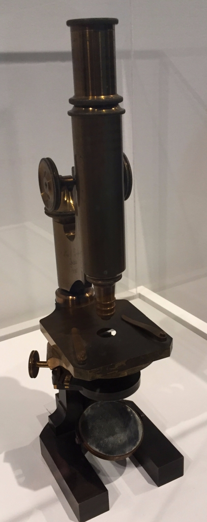
This modest microscope uses one eyepiece, one lens, and mirrored lighting. It was given to Cajal by the Spanish government in 1885, in recognition of his bacteriological studies during a cholera epidemic.
Cajal countered the reticular theory by asserting that there were spaces between neurons, the cells of the nervous system. Thus, with a very simple microscope (see above), Cajal saw gaps between cells in the nervous system, the key “observation” that led to the Neuron Doctrine. But the fact is that using this microscope, or even its modern descendants, the gaps between cells cannot be seen. They are simply not there. To visualize the gaps, an electron microsope, first introduced to neurobiology in the 1950s, is needed. Cajal reveals this in a pair of drawings (below) whose juxtaposition in The Beautiful Brain exhibit is pithily on point.


On the left side of the left image, you see a reticulated version of the spinal cord. The processes are all connected into a net. In contrast, the right image shows dense arbors that overlap but do not connect. This is the Neuron Doctrine in a nutshell. And the juxtaposition of these images shows that the switch came from Cajal’s brain and not from the brain sections.
Cajal and the Law of Dynamic Polarization
The second principle that Cajal is known for is the Law of Dynamic Polarization which states that information comes in through dendrites and leaves through an axon (axons away!) to contact another cell. Even with my appreciation that Cajal was inspired enough to see gaps where there were none visible, I marveled at Cajal’s deduction of physiology from slides. How did he figure out the direction of information flow within a neuron? You can’t see information, even with an electron microscope. I just did not get it. But now I do.
The answer revolves around the retina. Let me start by telling you that Cajal loved the retina. As he wrote in his autobiography:
“The retina is the oldest and most persistent of my laboratory loves. Life never succeeded in constructing a machine so subtly devised and so perfectly adapted to an end as the visual apparatus. I felt more profoundly than in any other subject to study the shuddering sensation of the unfathomable mystery of life.”
In the retina, Cajal could tell that the thick processes, dendrites, were directed toward the external world whereas the thin axons were directed to the brain. Okay. Simple. Logical. Observations of the olfactory bulb led to the same conclusion using similar logic. The exhibit states that the cerebellum also was central to Cajal’s conclusion that dendrites receive and axons send information (aka the Law of Dynamic Polarization) but the logic used here is not obvious to me.
Pathway drawings are quintessentially interpretative
Particularly exemplary of Cajal’s choice to interpret rather than document is his drawings of pathways. Obviously pathways are human constructions. They represent the shortest routes that we believe can join [every]here to [every]there. But our reification of these pathways does not require nature’s confirming participation. Thus pathways are constructions rather than objects visible under a microscope. I had not seen Cajal’s pathway drawings prior to attending the Beautiful Brain exhibit.
Cajal drew what would now be called a visual field pathway, which he deduced (correctly), ie that some axons from the retina cross and some do not, by imagining how an arrow, delightfully feathered, would hit the retina and be conveyed to the thalamus and in turn to visual cortex. He illustrated this in a style that is still used today even as his clear black and white drawing style has been replaced by [curmudgeon alert] busy pseudocolored graphics.
I particularly liked Cajal’s drawing of coughing and vomiting reflexes. These critical pathways are mostly ignored these days as neuroscientists obsess over an impoverished set of model movements (e.g. reaching). Yes, reaching is important – just ask anyone who can’t do it. But, no, reaching is not so important as to warrant the overwhelming majority of the motor control community working on this one problem. Cough is way cool for a lot of reasons which hopefully I will explore in a future post. Vomiting is also fascinating as it seamlessly combines autonomic and somatic control of smooth and voluntary muscles, respectively. Simply put, I like the non-cognitive side of life, represented beautifully by vomiting and coughing.
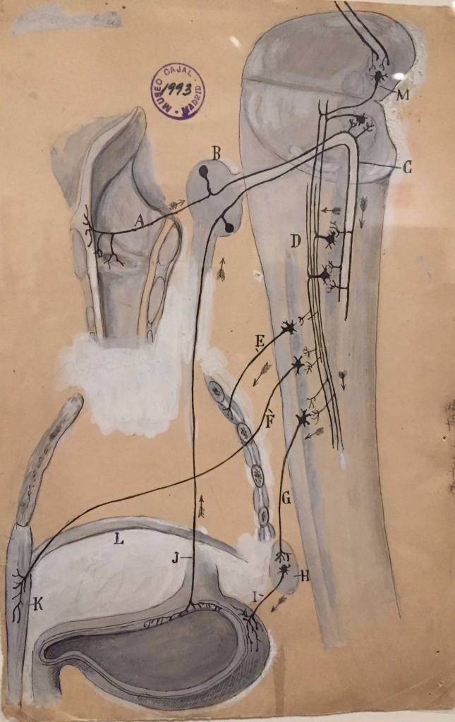
The cough reflex starts with irritation of sensory nerves from the larynx (A) that bring that irritating information to pre-motoneurons (M) that influence motoneurons (F) that control muscles of the chest and abdominal walls (K). The vomiting reflex is illustrated as starting with an irritation in the stomach that activates sensory nerves (J) that carry information to pre-motoneurons (D) that influence preganglionic motor neurons (G) that project to autonomic ganglionic neurons (H) which influence the smooth muscle of the stomach (I). The white in these drawings would not show up in the published versions.
Cajal did not draw every pathway correctly. For example, he drew motor pathways from motor cortex and cerebellum, incorrectly inking a direct projection descending from the cerebellar cortex to spinal motoneurons. [In fact, Purkinje cells project to deep cerebellar nuclei where neurons leave the cerebellum to synapse in hindbrain nuclei that in turn project to the spinal cord. So he missed a couple of neurons.]
The exhibit experience
Given the high degree of neuro-nerdiness intrinsic to this exhibit, I thought that I might run into people that I knew and indeed I did. I had barely gotten in the door when I saw Claire Daniele, a former UChicago postdoctoral fellow in Dan McGehee’s laboratory. Claire and I are even co-authors are on a paper. Claire had come in from Philadelphia with her husband specifically to see the exhibit. An hour or so later, a young woman asked timidly if I was Peggy Mason. She turned out to be a high school student from New Jersey who had taken my MOOC course: Understanding the Brain: The Neurobiology of Everyday Life.
The friendliness did not end there. The exhibit crowd was a highly self-selected group and the atmosphere reflected this. I highly recommend a visit to The Beautiful Brain. The exhibit is on through the end of March (March 31, 2018) at the Grey Gallery in New York City.
Concluding thoughts on the scientific method and reproducibility
We can learn from Cajal. He looked and looked at slides. He took in a great deal of data. But then he thought about it and drew what he constructed in his head. He did not try to take himself out of the equation. He was not attempting to reproduce what he saw when he looked down the scope of his microscope. He was not a slave to data.
Cajal’s drawings are not data. They are not reproducible. Cajal’s method should give us pause in our current uproar over reproducibility, accompanied by a complete devaluation of human impressions aka gut aka intuition aka speculative impressions.
I love data as much as the next modern scientist. But I also derive great value from impressions, those of my students and me as we do experiments, those of colleagues as they tell me about their work. I worry that we may only see the glancing spots of one of Cajal’s butterflies, ignoring them as they don’t reach significance:
“Like the entomologist in pursuit of brightly colored butterflies, my attention has chased, in the flower gardens of the gray matter, cells with delicate and elegant shpaes, the mysterious butterflies of the soul, the beatings of whose wings may some day – who knows? – clarify the secret of mental life.”
I conclude this post with a urge to put some art back into science, Sure, we can clearly label “speculation” as such but in the end I think we will all benefit if we let science out of its straightjacket.
Nerd alert!! Nerd alert!!
Below are things that I learned that rock my world because I am an unapologetic neuro-nerd. If you are not, you should stop reading here. If you are, dive in. Okay, here goes. Nerd-fest:
- Cajal’s drawings make it very obvious that the Purkinje cell has a recursive collateral that goes back into the Purkinje cell layer and even into the molecular cell layer. Never knew that. No idea what the import of that is.
- The narrative that I had always heard (and taught) was that once Golgi invented his eponymous method, Cajal used it to discover the neuronal world. But I learned that Cajal also used other stains such as the reduced silver nitrate method.
- People thought that dendiritic spines were artifacts. Cajal realized that they were not. One of the ways that he convinced himself of this was to show that spines were revealed by the Ehrlich methylene blue staining method as well as by the Golgi method.
- I believe there is one error in the exhibit. It is said that astrocytes are the most common glial cell in the brain. I always thought that was true but it turns out to be oligodendrocytes that are the most numerous.
- Speaking of errors, Cajal portrays inner hair cells with their hair bundles touching or minimally invested in the tectorial membrane while the hair bundles of the outer hair cells float free. Actually it is the opposite. I suspect some damage was done in the dissection, sectioning, or slide preparation.
- Apparently the sparrow has neurons in the brain that project into the inner nuclear layer of the retina. I have never heard of this. The retina, at least the mammalian one that I am aware of, has always stood out as a sensory system without brain-to-end-organ modulation. Is this brain-to-retina pathway prevalent among birds? Among reptiles? Is there any functional data?
- Cajal realized that the hippocampus was needed for olfactory memories. How did he figure that out?
- Cajal drew an amazing illustration of paracellular diapedesis. See below.

This shows a white blood cell moving out of a blood vessel (left) and into the tissue that the blood vessel serves. I am in awe at Cajal’s ability to put together a sensible (and correct from what I can tell) sequence from stills viewed in some haphazard order.
Categories: Book reviews, History, Science


I am so impressed with your cognitive and intuitive expressions, Peggy. You blend science and art so well. And, that’s the real beauty. This case here is an excellent example of STEAM, the ‘A’ being an essential part of STEM. I always believe that consummate learning is about integration and reification of data and ideas. On a lighter note I quote Einstein, “Reality is an illusion, albeit a persistent one.” Thanks for sharing your thoughts. I feel so nice to be your student.
LikeLike
Thank you Mahesh. All beautifully put. I feel so nice to be your friend.
LikeLike
I agre!! I´m started studying “Neurobiology” in the Coursera Plataform and I´m really impressed by how it´s posible to explain neurobiology in this rich way. Thank you so much, Dr. Peggy Mason (and sorry for my english)
LikeLike
I started*
LikeLike
I don’t know what your native language is but I’m very confident that your English is infinitely better than my ability in your language. Very happy that you’re enjoying the Coursera course.
LikeLike
After that fascinating post, I wish I could fly out tomorrow and visit Cajal’s exhibit. Truth be told, it makes me wish I was a member of the neuro-nerd club. What a cool world you inhabit.
LikeLike
Hi Lisa
How nice to hear from you. I think you are a neuro nerd. You are in my books anyway.
P
LikeLike
Aw thanks Peggy. That’s the best compliment ever. Hope you are well. I do y know if your aware but I started a new job at Northwestern working with a to of nerds from all stripes and disciplines. It’s a steep learning I g curve, buy they Are doing some family inatong stuff in the Chemistry of Life Processes Institue.
LikeLike
I found ur article so interesting and the drawing of the cells is is spectacular and I see the big cell at the bottom middle as a duck kicking up her feet and dancing. Thank you
LikeLike
Nice. Duck. Penguin. I saw it as a diving loon. I love you Merry. And it means the world to me that you stopped by.
LikeLike
Dear Peggy,
I really enjoyed reading this post!
Another anecdote from Cajal’s life is that he loved reading fiction. The obstacle was, that his father supposedly only allowed non-fiction books in their home library. His mother was the one who secretly kept some novels and opened the door to Cervantes, Hugo, etc…
Best,
Kirill
LikeLike
Thanks Kirill. Always wonderful to hear from you as always my friend. Cajal’s father sounds a bit unbearable. Glad that Cajal had the inner strength to forge his own path. At least in large part. Definitely good for us that Cajal’s father did not let him be an artist period stop.
LikeLike
I hope this exhibition comes to Brazil. I’m a designer who is now a PhD student in psychology / neuroscience and Cajal is a great inspiration to me. Btw, “I like the non-cognitive side of life, represented beautifully by vomiting and coughing” is a very quotable phrase!
LikeLike
That’s Karla!!!
LikeLike
New respect for Cajal and for you as ever. Thanks Peggy.
LikeLike
Thank you Francine!!
LikeLike
Dear Dr Mason
An other neuro-nerd here ;-)) and your student in Coursera. About “The retina, at least the mammalian one that I am aware of, has always stood out as a sensory system without brain-to-end-organ modulation” maybe you are interesting read this articles http://www.jneurosci.org/content/36/10/2904 and https://www.cmu.edu/bio/news/2016/kuhlman-neuronal-feedback.html
All the best,
Marta
LikeLike
Hi Marta.
Thanks for sending those articles. They both talk about cortical modulation back onto primary visual cortex. What Cajal drew was brain to retina. This is unknown in mammals.
I say visual system is unusual because there are projections from brain into vestibulum of inner ear and from spinal cord out to sensory nerve endings.
LikeLike
I just got my copy of this book and I love it. Thanks for beating me to blogging about it! Of course you also beat my by blogging about it competantly as opposed to what I’d have done. 🙂
LikeLike
Hey thanks almostrational!!!! Very nice words. I appreciate it.
P
LikeLiked by 1 person
Reblogged this on Almost Rational and commented:
This is the blog post I planned to right only much, much better!
LikeLike
Dear Dr Mason
I am your student in Coursera. I really enjoyed reading this post.
LikeLike
I’m so glad. Thanks for stopping by and I’m very happy that you’re interested in Neurobiology.
P
LikeLiked by 1 person
As a science loving artist, I really love schematics and visual representations of systems. And I love the idea of putting more science into art (rather than putting more art into science). Putting art into science seems like a good idea until the precision goes down and mistakes arise. That’s my only concern
LikeLike
Enjoyed reading your comments on this exhibit and excited to discover that these drawings are currently on display at the MIT Museum (not too far from me)! Just finished your Coursera course; the unit on movement/motor control and the last one on cognition were especially fascinating to me. But throughout the course I always appreciated your enthusiasm, insights, and “neuro-nerdiness” – great job!
LikeLike
Thanks so much. I had not heard that the show went to mit. That’s great. I didn’t know.
P
LikeLike